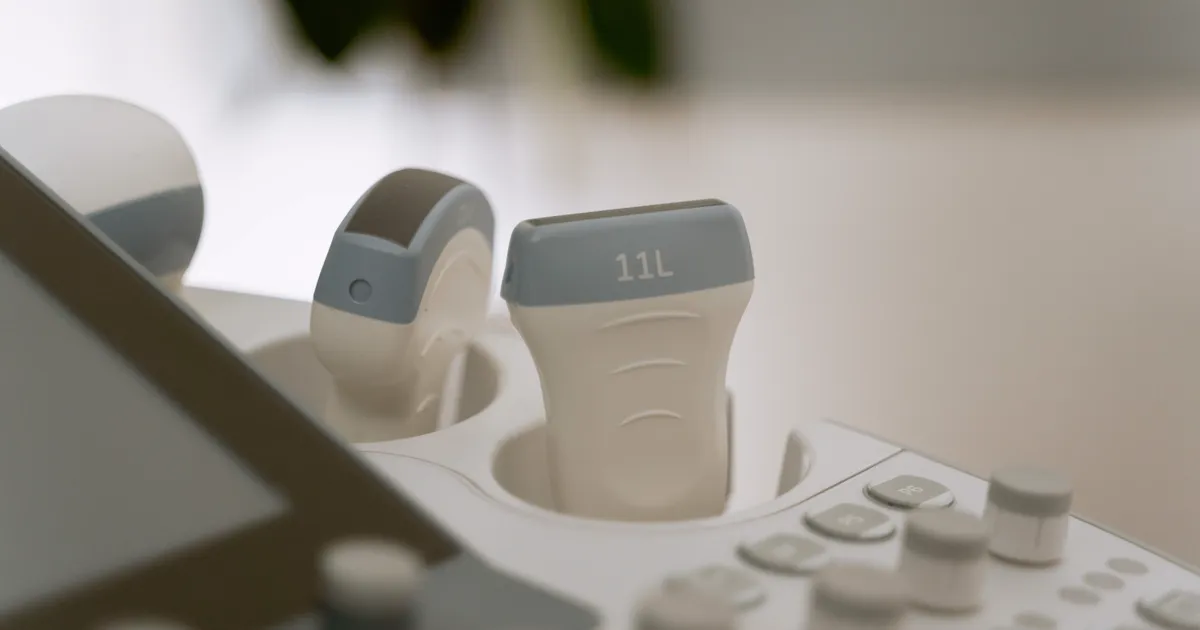
Assisted reproduction ends in a major congenital heart defect in 1.84% of births versus 1.15% after spontaneous conception — adjusted odds ratio 1.36 (95% CI 1.31–1.41), per a 2024 Nordic registry study of 7,747,637 liveborn children. After a normal anatomy scan, a screening fetal echocardiogram found nothing to treat in 91% of IVF pregnancies (Spurr et al., Pediatric Cardiology, 2022).
Should Every IVF Pregnancy Get a Fetal Echocardiogram?
No guideline recommends it firmly. The Society for Maternal-Fetal Medicine (SMFM) only suggests offering one — its weakest grade, 2C — and a 2025 comparison found assisted reproduction among the few risk factors where guidelines openly disagree.
“We suggest that fetal echocardiography be offered to patients with pregnancies achieved with in vitro fertilization and intracytoplasmic sperm injection (GRADE 2C).” — SMFM Consult Series #60, American Journal of Obstetrics & Gynecology, 2022
GRADE 2C means two things: “we suggest,” not “we recommend,” and “very low certainty” evidence. The same 2022 SMFM consult grades the detailed anatomy ultrasound 1B — firm, on moderate-quality evidence — so clinicians treat the anatomy scan, not the echo, as the test that matters.
The 2018 meta-analysis said it plainly: “There is no consensus in current practice guidelines on whether conception by IVF/ICSI techniques is an indication for performing a fetal echocardiogram” (Giorgione et al., 2018).
Data comes from SMFM Consult Series #60, the CoNARTaS Nordic registry study, the 2025 guideline comparison, the 2021 Duke cost model, the 2022 Seattle yield study and CDC heart defect data, compiled by the ProIVF Medical Editorial Team and reviewed by the ProIVF Medical Advisory Board.
What Did the 2025 Guideline Comparison Find?
The guideline comparison (De Robertis et al., 2025) weighed 17 congenital-heart-disease risk factors across six English-language guidelines (2008–2023): only 3 won complete agreement.
3 of 17 (17.6%) had complete agreement — all three fetal findings: suspected heart defect at the anomaly scan, a major extracardiac abnormality, non-immune hydrops fetalis. 8 of 17 (47.1%) had partial agreement: family history, increased nuchal translucency, multiple pregnancy, maternal diabetes, teratogen exposure.
The remaining 6 of 17 (35.3%) produced complete disagreement — including “use of assisted reproductive technology,” fetal genetic anomalies and first-trimester sonographic markers. The review’s authors concluded that “areas of controversy were greater in quantity than areas of consensus.”
What Is a Fetal Echocardiogram, and How Does It Differ from the Anatomy Scan?
A fetal echocardiogram is a dedicated ultrasound of the fetal heart, read by a sonographer or cardiologist; the anatomy scan surveys the whole baby, heart included.
| Detailed anatomy ultrasound | Fetal echocardiogram | |
|---|---|---|
| What it covers | All organ systems, growth, placenta, amniotic fluid, cord insertion | The heart only: chambers, valves, great arteries, rhythm, blood flow |
| Typical window | 18 0/7–22 6/7 weeks (SMFM’s anatomy-scan window) | Booked after the anatomy scan; earlier in specialized centers |
| Who reads it | Obstetrician, maternal-fetal medicine specialist or radiologist | Pediatric or adult congenital cardiologist, or a specialized sonographer |
| Extra views | Four-chamber and outflow-tract views at screening level | Sequential segmental analysis, Doppler, three-vessel trachea view, measurements of every structure |
| Cost logic in the IVF model | Base-case average $8,119 per patient | Universal echo for all IVF pregnancies raised the average to $8,560 |
The cost row comes from the 2021 Duke model: three screening policies through one year after delivery.
What Does the Sonographer Actually Look At?
The minimum useful screening set is the four-chamber view plus the two outflow tracts — the Seattle study’s definition of a “complete” obstetric scan (Spurr et al., 2022).
Supplemental views (three-vessel, sagittal aortic arch) changed nothing, and accuracy did not differ by reader specialty: after a normal obstetric scan, the echo was also normal in 95% of obstetrician, 85% of maternal-fetal medicine and 92% of radiology reads (p = 0.95).
Is a Fetal Echocardiogram Worth It After a Normal Anatomy Scan?
Usually little: of 304 IVF pregnancies with normal cardiac anatomy on the anatomy scan, 278 (91%) were also normal on screening echocardiography.
Of the 26 abnormal results, none required intervention — 17 muscular and 5 perimembranous ventricular septal defects, 4 minor valve abnormalities. The authors concluded that “consideration should be given to the cost/benefit of screening F-echo for the indication of IVF if normal cardiac anatomy is demonstrated on OB-scan.”
How Much Higher Is the Heart Defect Risk After IVF?
Three large studies agree: the excess is small in each — at most 1.84% versus 1.15%.
| Study | Population | Defects after IVF/ART | After spontaneous conception | Effect size |
|---|---|---|---|---|
| CoNARTaS, European Heart Journal, 2024 | 7,747,637 liveborn children, Denmark/Finland/Norway/Sweden; 171,735 after ART | Major CHD 1.84% (3,159 children) | 1.15% (86,824) | Adjusted OR 1.36 (1.31–1.41) |
| Same study, severe defects | Same cohort | Severe CHD 0.35% (594) | 0.26% (19,375) | Adjusted OR 1.30 (1.20–1.42) |
| Giorgione meta-analysis, 2018 | 8 cohorts: 25,856 IVF/ICSI vs 287,995 spontaneous | Total CHD 1.30% (337) | 0.68% (1,952) | Pooled OR 1.45 (1.20–1.76); singletons 1.55 (1.21–1.99) |
| Appiah, Human Reproduction, 2024 | 9.6 million US singleton livebirths to first-time mothers, 2016–2022 | Cyanotic CHD (CCHD) 1.2 per 1,000 (ART 1.1; non-ART fertility treatment 1.3) | 0.5 per 1,000 | Adjusted OR 2.06 (1.82–2.33); ART alone 2.02 (1.73–2.36) |
What Do Those Percentages Mean in Real Numbers?
In the largest study, 1.84% is roughly 1 in 54 IVF-conceived children; 1.15%, about 1 in 87 spontaneously.
The gap is roughly 7 additional major heart defects per 1,000 births; for severe defects, 0.35% versus 0.26% is about 1 extra per 1,100 births. Nearly nine in ten IVF-conceived children had no major defect; the study stresses “the absolute risks are, however, modest.” In the US cyanotic-defect cohort, overall prevalence was 0.6 per 1,000 live births across 5,287 cases, and the share of births following infertility treatment had risen from 1.9% in 2016 to 3.1% in 2022 (Appiah et al., 2024).
Are Twins the Real Reason?
Partly: in the CoNARTaS cohort of 171,735 ART-born children, multiples carried the highest risk “regardless of conception method,” the authors report.
The same study found no meaningful difference between ICSI and conventional IVF, or between frozen and fresh transfer (Sargisian et al., 2024) — the lab technique does not separate the groups. A US cohort agrees: cyanotic defects were equally common after non-ART fertility treatment (1.3 per 1,000) and ART (1.1 per 1,000) (OR 1.04, 0.82–1.33).
Two explanations remain: the infertility itself (with parental age and health), and the prematurity and low birthweight that IVF singleton and twin pregnancies carry. If transferring one embryo, the elective single embryo transfer discussion matters more.
Does PGT-A, ICSI or Donor Eggs Change the Number?
Preimplantation genetic testing for aneuploidy (abnormal chromosome counts) screens chromosome number, not heart structure: a euploid (chromosomally normal) transfer does not lower the 1.84% figure.
SMFM still advises offering prenatal genetic screening and diagnostic testing “regardless of whether preimplantation genetic testing has been performed” (GRADE 1C); see our PGT-A explained guide.
What Does Universal Fetal Echocardiography Cost?
The Duke model priced three IVF policies; the two adding a routine echocardiogram were poor value (Chung et al., 2021).
| Screening strategy | Average cost per patient | Effectiveness (QALYs) | Incremental cost per extra QALY |
|---|---|---|---|
| Anatomy ultrasound, echo only if the heart looks abnormal | $8,119 | 1.74487 | — |
| Echo for every IVF pregnancy with ICSI | $8,408 | 1.74497 | $2,840,494 |
| Echo for every IVF pregnancy | $8,560 | 1.74499 | $5,692,457 |
Both ratios far exceed the standard willingness-to-pay threshold of $50,000–$100,000 per quality-adjusted life year (QALY). For major defects alone, echoing every ICSI pregnancy cost $527,562 per additional defect detected and every IVF pregnancy $790,510 per additional case — about five times more to find one extra major defect than a newborn’s first year of care costs, the authors note.
Their conclusion: “The most cost-effective method of screening for CHDs in pregnancies following IVF … is to perform a fetal echo only when abnormal cardiac findings are noted on the detailed anatomy scan. Performing routine fetal echo for all IVF pregnancies is not cost-effective.”
Who Pays for the Scan?
Your insurer decides: the echo is billed separately from the anatomy ultrasound, and in the US coverage depends on whether the interpreting cardiologist is in network.
Ask three questions first: the referral’s coding (screening or diagnostic), your imaging deductible and coinsurance, and whether a pediatric cardiology consult is extra. None of this changes the clinical decision; it changes the bill.
When Is a Fetal Echocardiogram Clearly Worthwhile?
All six guidelines agree on 3 indications — all three fetal findings; several maternal ones are common enough in IVF populations to matter.
- A suspicious heart on the anatomy scan — one of only three complete-agreement indications (De Robertis et al., 2025).
- A major abnormality outside the heart, or non-immune hydrops — the other two universally agreed indications.
- A multiple pregnancy — partial agreement; the risk group CoNARTaS singles out regardless of conception method.
- Increased nuchal translucency or an abnormal first-trimester screen — partial agreement; see our genetic screening and PGT guide.
- Maternal diabetes, PKU (phenylketonuria), autoimmune disease or teratogen exposure — partial-agreement indications, defined inconsistently across guidelines.
- A family history of congenital heart disease, or a sibling with one — partial agreement, and the one insurers accept without argument.
What Else Does SMFM Recommend Differently for IVF Pregnancies?
Ten of the twelve recommendations in Consult Series #60 are not about the heart at all.
| Recommendation | Grade |
|---|---|
| Offer genetic counseling to all IVF/ICSI patients | 2C |
| Offer prenatal genetic screening and diagnostic testing (CVS or amniocentesis) even after PGT | 1C |
| Discuss the accuracy of first-trimester screening, including cfDNA | 1A |
| Counsel about multifetal pregnancy reduction if multiples occur | 1C |
| Detailed anatomy ultrasound (CPT 76811) for IVF/ICSI pregnancies | 1B |
| Offer fetal echocardiography | 2C |
| Assess placental location and shape, cord insertion, vasa previa (vessels running across the cervical opening) | 1B |
| No routine serial cervical length | 1C |
| Third-trimester growth assessment at least once, no serial scans for IVF alone | 2B |
| No low-dose aspirin for IVF as the sole preeclampsia indication | 1B |
| Weekly antenatal fetal surveillance from 36 0/7 weeks | 2C |
| Shared decision-making about induction at 39 weeks | 1C |
Two rows surprise patients: induction at 39 weeks is a shared decision, not a rule, and IVF alone warrants no aspirin or serial growth scans. What IVF reliably changes is the monitoring calendar — and the odds of twins, where most risk concentrates.
If a Heart Defect Is Found, What Happens Next?
Most prenatal findings are minor: 3 in 4 babies with a heart defect do not have the critical kind.
The US Centers for Disease Control and Prevention reports that “heart defects affect nearly 1% of births, or about 40,000 babies, each year” and that “about 1 in 4 babies born with a heart defect has a critical heart defect,” needing “surgery or other procedures in the first year of life.” The other three in four do not.
All 26 defects the Seattle screening found were the non-critical septal and valve abnormalities above — none required intervention.
Can It Be Detected Before Birth, and Does That Matter?
“Some heart defects can be found during pregnancy using a special type of ultrasound called a fetal echocardiogram. Other heart defects aren’t detected until birth or later in life,” CDC states.
The echo is a screening tool with partial detection, not a guarantee. Where a critical defect is found before birth, the benefit is planning: delivery hospital, on-site pediatric cardiology, and the post-birth confirmatory-echo-and-treatment sequence. All US newborns then get pulse-oximetry screening, catching what prenatal imaging missed.
Patient Stories: Three IVF Pregnancies, Three Different Choices
Patient stories are shared with consent; names and identifying details changed; clinical figures reflect the sources cited above.
Case 1: “The clinic said it was standard, so I just had it” — Kate, 34, Toronto
Kate’s single embryo transfer at a Toronto clinic cost about CA$14,000 all-in — a singleton pregnancy, no other risk factors. Her 20-week anatomy scan was normal, but the clinic ordered a fetal echocardiogram anyway under its IVF protocol — no out-of-pocket charge under provincial coverage.
“Nobody explained to me that it was a ‘suggestion, very low certainty’ rather than a rule,” she says. The echo was normal; her daughter was born healthy at 39 weeks.
Case 2: “We paid for it and it changed nothing” — Nadia, 38, Dubai
Nadia’s two IVF cycles in Dubai cost roughly AED 92,000 including medications. At 22 weeks in a twin pregnancy, the obstetric scan flagged one twin’s outflow tract as poorly visualized, so they were referred for a fetal echocardiogram at a private cardiology center.
Both hearts were normal; delivery was planned at a hospital with a neonatal intensive care unit — a decision driven by the twins, not the echo. The scan cost a few hundred dollars more than the anatomy ultrasound.
“It bought us an appointment with someone who could answer questions, more than it bought us information,” she says. Both boys were born at 35 weeks and went home together at 38 weeks.
Case 3: “Our baby had a defect — and the anatomy scan had already seen it” — Elena and Marco, 32 and 35, Milan
Elena conceived after three IVF attempts at a Milan clinic, about €9,500 total, including Italian medications. At the 21-week anatomy scan the sonographer identified an atrioventricular septal defect and referred them to a fetal cardiology unit; the echocardiogram confirmed it and mapped the plan.
Their daughter was born at a hospital with pediatric cardiac surgery and underwent repair at four months. “The extra scan did not find the problem — the anatomy scan did,” Marco says. “What the echocardiogram gave us was the surgeon, the delivery plan and six months of preparation instead of six hours.”
FAQ
Q: Is a fetal echocardiogram the same as the 20-week anatomy scan?
No. The anatomy scan is one study, done between 18 0/7 and 22 6/7 weeks, that surveys every organ system and includes screening heart views—the four-chamber view and the 2 outflow tracts.
A fetal echocardiogram is a dedicated cardiac exam with Doppler measurement of every structure.
Q: Why do some clinics order a fetal echocardiogram for every IVF pregnancy?
SMFM only “suggests” it (GRADE 2C), so practice varies by country, clinic protocol and payer. The 2025 guideline review placed assisted reproduction among the completely disputed indications (6 of 17 risk factors, 35.3%).
Q: My anatomy scan was normal. What is the chance the echocardiogram finds something?
In the largest study of this question, 9% of screening echocardiograms after a normal obstetric scan were abnormal (26 of 304 IVF pregnancies), and none of those 26 required intervention (Spurr et al., 2022).
Q: Does IVF cause heart defects?
No causal mechanism has been established. The CoNARTaS authors attribute part of the excess to multiple pregnancies; a US cohort found cyanotic defects equally common after ART (1.1 per 1,000) and non-ART treatment (1.3 per 1,000) — pointing to infertility, not the lab.
Q: What share of babies born after IVF actually has a heart defect?
In the largest Nordic cohort about 1.84% — roughly 1 in 54 — have a major heart defect, versus 1.15% (about 1 in 87) after spontaneous conception; severe defects run 0.35% versus 0.26%.
Q: Is the extra scan worth it for a twin pregnancy?
Twice as many hearts — and multiples carry the highest heart-defect risk in CoNARTaS, “regardless of conception method.” Twins bring other monitoring too: SMFM recommends counseling about multifetal pregnancy reduction (GRADE 1C) and weekly surveillance from 36 0/7 weeks (GRADE 2C).
Q: Will insurance cover it?
Coverage depends on your plan, the referral’s coding (screening or diagnostic) and whether the interpreting cardiologist is in network. The figures above ($8,119 to $8,560 per patient) model societal costs — imaging, consults, surgery, caregiver productivity loss — not your bill.
Q: If we transfer one embryo, do we still need to think about this?
Then you have removed the largest share of the excess: CoNARTaS heart-defect risk “was highest in multiples, regardless of conception method.”
For a singleton the increase is real but small (Giorgione et al., 2018: OR 1.55, 95% CI 1.21–1.99, against a 0.68% baseline). With an uncomplicated singleton and a normal anatomy scan, the 91% normal-yield figure makes a fetal echocardiogram optional rather than necessary — and if you conceive abroad, ask which of the 12 SMFM recommendations the clinic’s prenatal pathway follows.
How to Have This Conversation With Your Clinician
Three questions settle most of it:
- “What would change if the echocardiogram is normal — and what changes if it is not?” If the answer is “nothing,” the test is reassurance, and reassurance has a price of its own.
- “Were the four-chamber and outflow-tract views obtained on my anatomy scan?” A complete study with those views is the strongest reason to skip a screening echo; an incomplete study is the clearest reason to have it.
- “Would you order this for a spontaneously conceived pregnancy with the same history?” This isolates whether IVF alone is driving the referral, or something specific to you.
Compare clinics through verified hospital profiles, read how IVF affects pregnancy timing and outcomes, or contact our team about a specific clinic’s protocol.
About this article: Researched and written by the ProIVF Medical Editorial Team from the SMFM (2022), CoNARTaS (2024), Giorgione (2018) and Appiah (2024) studies, the 2025 guideline comparison, the 2021 Duke cost model, the 2022 Seattle yield study and CDC data, with AI tools used only for literature organization and initial drafting under editorial direction. Reviewed by the ProIVF Medical Advisory Board; our editorial standards are on our About page.
Last updated: October 15, 2026. Informational only, not medical advice: prenatal-imaging decisions belong to you and your obstetric or fetal-medicine team, who know your history; population statistics cannot predict an individual pregnancy.